DETERMINATION OF SHAPES, DIMENSIONS AND COLOURS OF SPORES, LAYERS, AND MYCORRHIZAE
 |
 |
 |
 |
 |
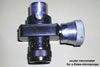 |
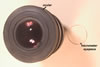
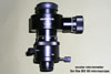
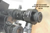
The shape and size of spores characterized here were determined based on at least 50 intact spores mounted in a drop of water or lactic acid placed on a microscope slide. The dimensions were determined using a light microscope equipped with an ocular micrometer.
The thickness of layers of spore wall and germination walls was measured in spores freshly isolated and crushed in PVLG or PVLG+Melzer’s reagent (1:1, v/v) and observed under a light microscope equipped with a micrometer eyepiece. Storage of spores in, e. g., lactic acid usually makes layers to thicken and changes their staining intensity in Melzer’s reagent.
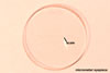
The measurements of mycorrhizae were made using a light microscope equipped with an ocular micrometer or a micrometer eyepiece.
 |
 |
 |
 |
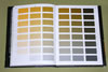
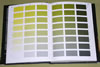
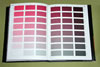
Colours of mycorrhizae come from roots stained in 0.1% trypan blue, mounted in PVLG, and observed under a light microscope.
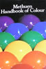
Colours were determined according to Kornerup and Wanscher (1983).
REFERENCE
Kornerup A., Wanscher J. H. 1983. Methuen handbook of colour. 3rd Ed. E. Methuen and Co., Ltd., London. 252 pp.