GERMINATION.
Not recognized to date.
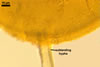
MYCORRHIZAE.
Glomus spinuliferum has originally been found associated with mycorrhizal roots of plants of a semi-natural grassland (Oehl et al. 2003). All attempts to grow this fungus in both trap and one-species cultures failed.
DISTRIBUTION.
The holotype of Gl. spinuliferum came from spores isolated from a soil sample originating from a semi-natural grassland located at Vogelsang Pass near Vogtsburg, belonging to the Kaiserstuhl Nature Reserve, Germany. The only other report of this fungus is that of Oehl et al. (2005) informing of the presence of its spores in soils of grasslands and a vineyard located in the Upper Rhine Valley between Basel (Switzerland), Freiburg i. Br. (Germany), and Mulhouse (France).
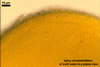
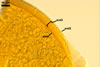
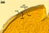
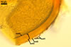
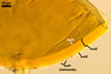
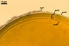
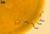
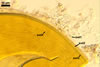
NOTES. Apart from Gl. spinuliferum, other species producing spores ornamented with spines projecting outward are Gl. monosporum Gerd. & Trappe, Gl. pansihalos S.M. Berch & Koske, and Gl. spinosum H.-T. Hu. Compared with Gl. spinuliferum, spores of Gl. monosporum are almost 2-fold larger [140-330 µm vs. (110-)120-140(-154) µm diam in Gl. spinuliferum] and their wall consists of only two layers (Gerdemann and Trappe 1974), and not four ones. Additionally, the distribution of spines on spores of Gl. monosporum is less both uniform and compact. Finally, spores of Gl. monosporum occur in sporocarps, whereas Gl. spinuliferum forms only single spores.
Glomus pansihalos, the most morphologically similar species to Gl. spinuliferum, differs from the fungus compared here mainly in colour of its spores, as well as in size and the density of the processes ornamenting the spores. Spores of the former species may be darker [up to dark yellow (4C8) vs. up to orange (5A7) in Gl. spinuliferum], and their processes are both lower (1x1x1 µm vs. 1-2(-3) x 1.5 µm in Gl. spinuliferum) and more widely dispersed (1-5 µm vs. no space or space smaller than 1 µm in Gl. spinuliferum; Berch and Koske 1986; Błaszkowski 2003).
Glomus spinuliferum is easy to distinguish from Gl. spinosum, because the latter species forms spores both much darker (orange-brown to nearly black vs. sunflower yellow to orange in Gl. spinuliferum) and smaller [40-90 µm diam vs. (110-)120-140(-154) µm diam; Hu 2002]. Additionally, spores of Gl. spinosum are born in sporocarps, and those of Gl. spinuliferum occur only singly in the soil.
REFERENCES
Berch S. M., Koske R. E. 1986. Glomus pansihalos: a new species in the Endogonaceae, Zygomycetes. Mycologia 78, 838-842.
Błaszkowski J. 2003. Arbuscular mycorrhizal fungi (Glomeromycota), Endogone and Complexipes species deposited in the Department of Plant Pathology, University of Agriculture in Szczecin, Poland. http://www.agro.ar.szczecin.pl/~jblaszkowski/.
Gerdemann J. W., Trappe J. M. 1974. The Endogonaceae in the Pacific Northwest. Myc. Memoir 5, 1-76.
Hu H.-T. 2002. Glomus spinosum sp. nov. in the Glomaceae from Taiwan. Mycotaxon 83, 159-164.
Oehl F., Wiemken A., Sieverding E. 2003. Glomus spinuliferum, a new ornamented species in the Glomales. Mycotaxon 86, 157-162.
Oehl F., Sieverding E., Ineichen K., Ris E.-A., Boller T., Wiemken A. 2005. Community structure of arbuscular mycorrhizal fungi at different soil depths in extensively and intensively managed agroecosystems. New Phytol. 165, 273-283.