
DETERMINATION OF SHAPES, DIMENSIONS, AND COLOURS OF FUNGAL STRUCTURES
|
|
|
|
|
|
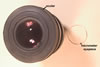
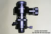
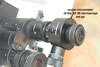
When investigated personally, the shape and size of all the structures characterized in the website presented here were determined based on at least 50 specimens mounted in a drop of water, lactic acid, polyvinyl alcohol-lactic acid-glycerol (PVLG), or PVLG+Melzer’s reagent (1:1 v/v) placed on a microscope slide. The dimensions were determined using a light microscope equipped with a micrometer eyepiece or an ocular micrometer.
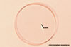
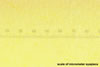
The thickness of layers of spore wall and germination walls of arbuscular mycorrhizal fungi of the phylum Glomeromycota was measured in spores freshly isolated and crushed in PVLG or PVLG+Melzer’s reagent. Storage of spores in, e. g., lactic acid usually makes layers to thicken and changes their staining intensity in Melzer’s reagent. The thickness of spore wall layers and germination wall layers (in Archaeospora, Acaulospora, Entrophospora, Pacispora, and Scutellospora spp.) was determined under a light microscope equipped with a micrometer eyepiece.
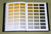
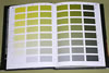
Colours of the structures examined were determined when they were in water, lactic acid, or PVLG, as well as in PVLG+Melzer’s reagent when staining reactions were to be examined.
 |
 |
 |
 |
Colours of mycorrhizae come from roots stained in 0.1% trypan blue, mounted in PVLG, and observed under a light microscope.
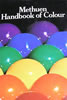
Colours were determined according to Kornerup and Wanscher (1983).
REFERENCE
Kornerup A., Wanscher J. H. 1983. Methuen handbook of colour. 3rd Ed. E. Methuen and Co., Ltd., London. 252 pp.