ISOLATION OF SAPROTROPIC AND PATHOGENIC FUNGI FROM PLANT ORGANS AND CULTIVATION OF THE FUNGI IN AGAR CULTURES
When structures of a fungus were visible on plant organs during their collection, agar cultures of the fungus were initiated from them. Using a dissecting microscope, the structures (spores, mycelium, etc.) were transferred on the surface of the agar medium used by means of the end of a sterile needle and then grown at a room temperature. If more than one organisms appeared on the medium, one-species cultures of each of them were established.
 |
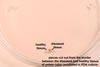 |
When diseased tissues of the plant organ examined lacked any visible fungal structure, small, ca. 3 x 3 mm, pieces were first aseptically cut out from the border between the diseased and healthy tissue and then placed on the surface of the medium used (Baudoin et al. 1988).
 |
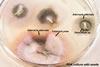
Alternatively, the plant organ with diseased symptoms was incubated in a moist chamber to initiate the formation of any structure of the putative fungus. Moist chambers were earlier autoclaved Petri plates containing a piece of filter paper to which sterile water was added to wet the paper. Then, infected plant material was placed in the chamber and incubated under a light at a room temperature for 1-3 days. When a fungus appeared, it was isolated and established in a pure culture, as described above. Establishment of moist chamber also is a good way to obtain fungal spores for identifying the pathogen.
Saprotrophic and pathogenic fungi of plant organs with no disease symptoms, e. g., seeds, were isolated after incubation of the organs in 10-cm Petri dishes with potato dextrose agar (PDA) + 100 µg/ml streptomycin sulfate for 10-14 days at a room temperature. Before the incubation, the organs were usually surface disinfested by their immersion in 0.1% HgCl2 in 10% CH4OH for 30 second, then washed in three changes of sterile water, and finally dried between two sheets of a sterile filter paper. Surface disinfestation may also be done in a NaOCl solution containing 1% chlorine for 10 min.
 |
 |
 |
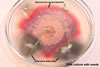 |
1-day old |
5-day old |
7-day old |
|
Sporulation of most of the fungi examined stimulated their lighting with cool day-light-type white fluorescent lamps positioned ca. 30 cm above the plant organs incubated. The time of lighting ranged from 12 to 16 h per 24 h.
 |
REFERENCES
Baudoin A. B. A. M., Hooper G. R., Mathre D. E., Carroll R. B. 1988. Laboratory execrcises in plant pathology: an instructional kit. APS Press. The Phytopath. Soc. St. Paul, Minnesota.
Dhingra O. D., Sinclair J. B. 1985. Basic plant pathology methods. CRS Press, Inc. Boca Raton, Florida.
Neergaard P. 1977. Seed pathology. The Macmillan Press Ltd. London.